Robotics and Intraoperative Imaging in Spine and Cranial Surgery

Surgical robotics is often misunderstood. In spine and cranial surgery the robot does not replace the surgeon. It holds a planned trajectory with a steadiness and repeatability that a human hand cannot sustain, so the surgeon's judgment is executed more precisely. The decision stays human, the execution gets steadier.
Alignment and imaging together
A modular robotic arm such as Brainlab Cirq auto-aligns to the planned trajectory, and a bone-anchored drill guide helps prevent the instrument from skiving off course during screw placement. The robot is only as good as the plan and the image it works from, which is why imaging is the other half of the story.
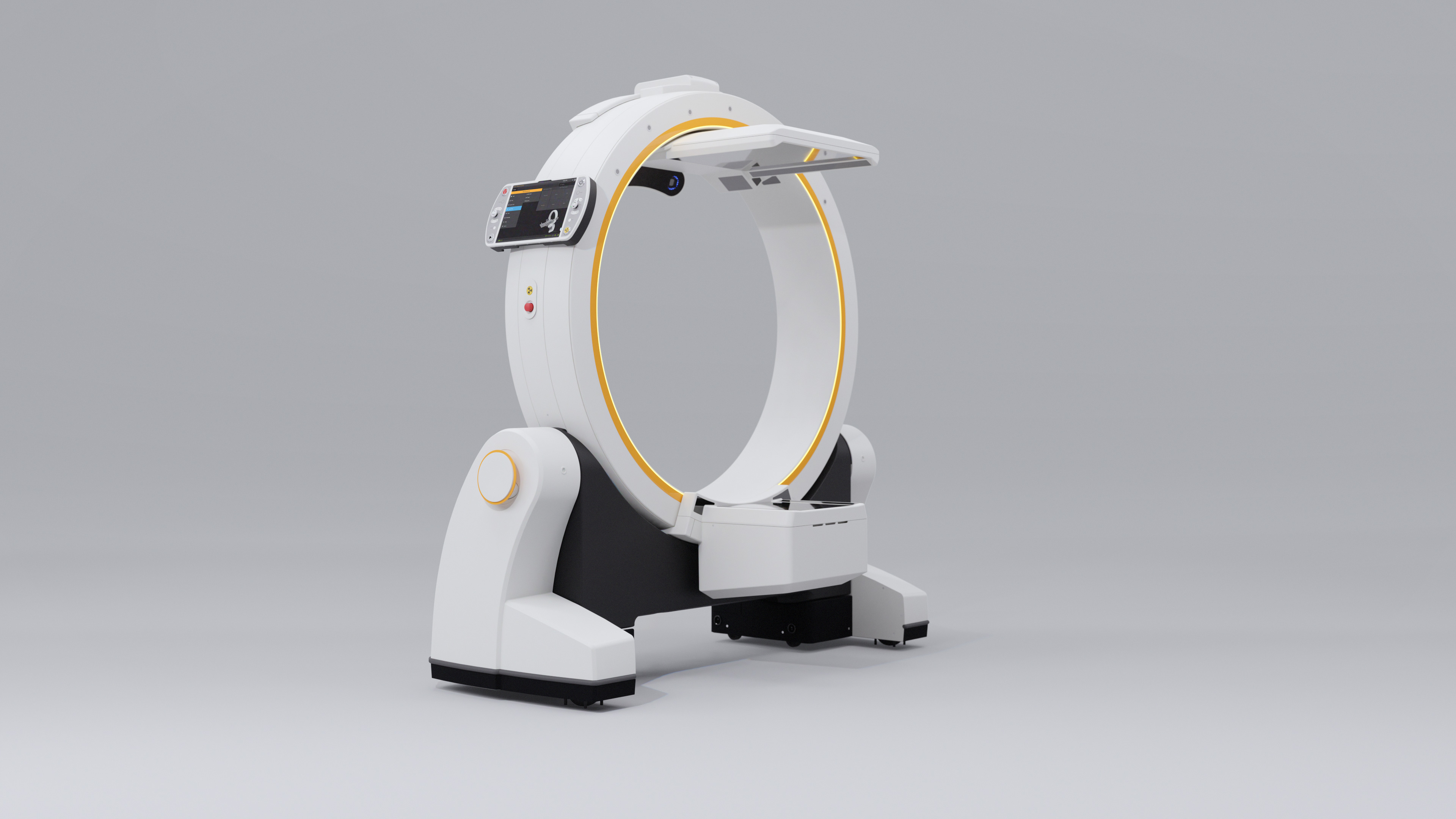
A mobile imaging robot like Loop-X delivers intraoperative cone-beam CT and registers the patient to navigation automatically, so the plan, the image and the robotic guidance share one coordinate space. When all three agree, the team works with a level of confidence that is hard to reach with any one of them alone.
Seeing the plan in space
Mixed reality adds another layer. Patient-specific anatomy rendered as interactive 3D, reviewed before surgery and brought into the room, helps the surgeon hold a spatial sense of the plan that a flat screen cannot give. It is most useful in complex cranial and spine work where orientation is everything.

The practical case
Because these platforms are designed to be modular and vendor-neutral, a hospital can add robotics and imaging to a navigated room rather than committing to a closed system. Brainz views robotics and imaging as reinforcements to the planning and navigation backbone, not standalone purchases. The benefit is greatest when every element acts on the same plan.
Product availability, indications, regulatory status and clinical use vary by country and follow manufacturer documentation and applicable regulatory approvals. Trademarks belong to their respective owners.