A hip or knee program is judged on what happens between the first X-ray and the last implant check. Most of the risk sits in the handoffs: from image to plan, from plan to table, from table to confirmation. When each handoff belongs to a different system, details are lost. When they belong to one pathway, the plan survives contact with the operating room.
Step one: the plan lives on the image
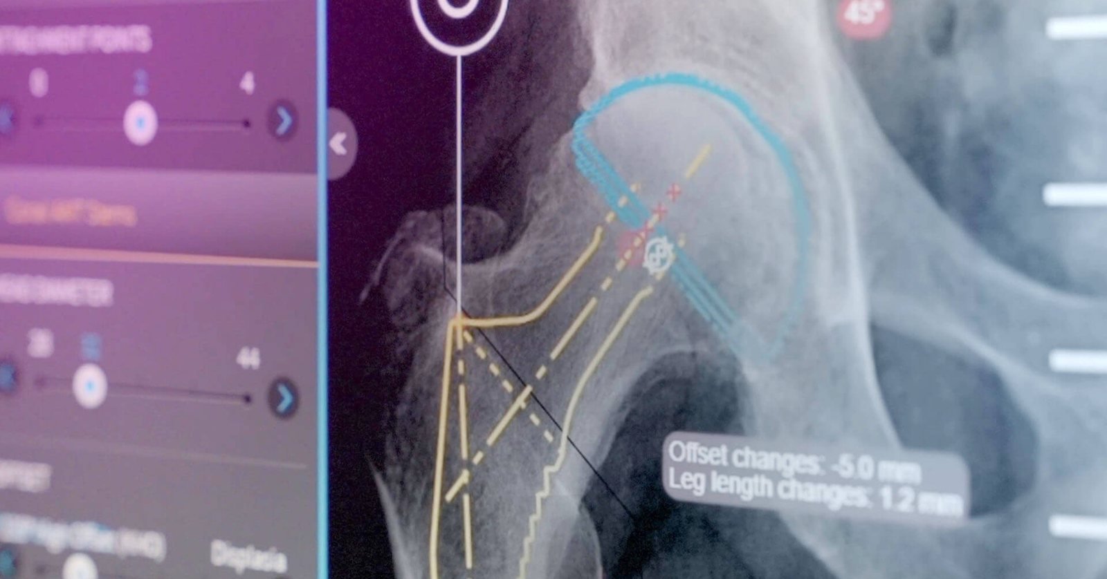
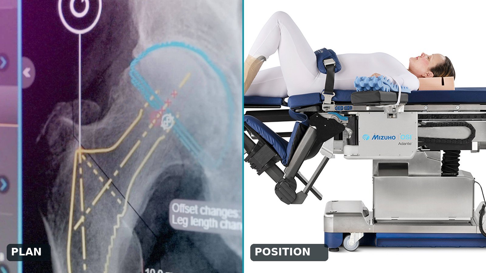
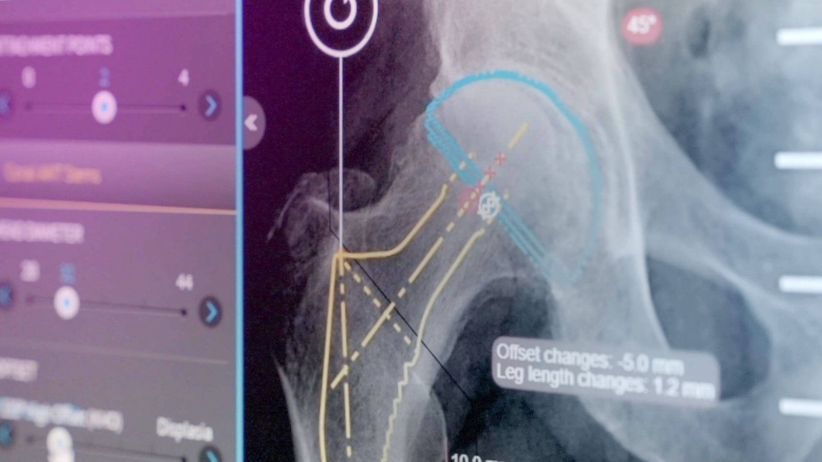
The pathway starts with Brainlab TraumaCad. The surgeon overlays implant templates on the calibrated X-ray and decides size, offset and alignment before the patient is booked. The platform covers hip, knee, trauma and deformity cases and carries a library of roughly 4,000 implant templates, so the plan reflects the implants the hospital actually stocks. Because it integrates with PACS, the approved plan is saved to the record and can be opened by anyone on the team.
Step two: position the patient for the plan
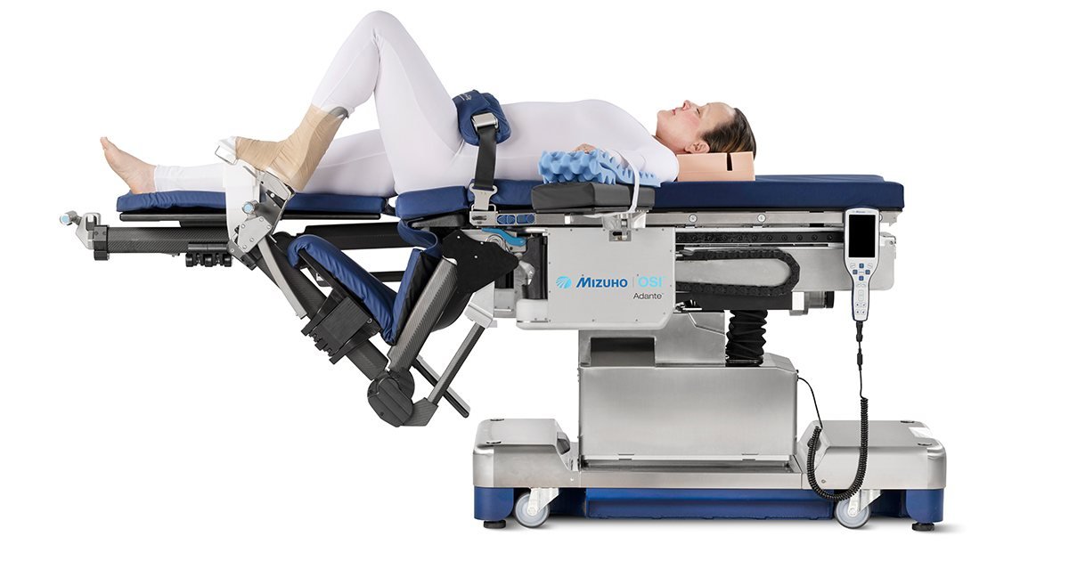
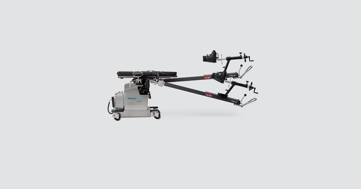
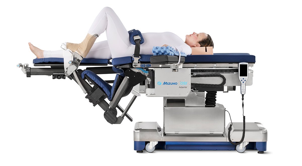
A plan only helps if the table lets the team execute it. For anterior approach hip work, the Mizuho OSI Hana table positions each leg independently and applies traction, with a radiolucent structure that keeps fluoroscopy views clean. For a mixed joint list, Mizuho OSI Adante extends the idea to the knee. Its FlexLation Spar moves the knee from full extension to deep flexion with a position and hold action, and a quick foot release frees the foot for soft tissue and balance assessment. On the hip side, Femur Lift exposes the resected femur during the anterior approach, and the platform keeps unimpeded C-arm access for lower limb imaging.
Step three: confirm in the room, not after it
Intraoperative fluoroscopy is the standard way to check cup position and leg length in the direct anterior approach, and the question now is how much of that check can be measured rather than judged by eye. A 2025 study in Cureus compared 38 patients whose direct anterior hips were checked with software-aided fluoroscopy against 38 controls checked with fluoroscopy alone. Mean postoperative leg length discrepancy was 2.2 mm with software against 4.6 mm without, and every hip in the software group (39 of 39) was within 10 mm, against 37 of 38 in the control group.
The study is small, and its control group was retrospective, so it is a signal and not a verdict. The software it tested is a separate tool, not part of the Brainz portfolio. The point for a program is the workflow: a plan that carries a numeric target, a radiolucent table that keeps the image clean, and a measured comparison while the patient is still on the table.
Step four: close the loop
The pathway finishes where it began, on the image. The postoperative X-ray is compared with the TraumaCad plan, and the difference becomes data the team can review. Over a year, that comparison shows whether sizing habits, positioning routines or table setups need to change. A program that records the gap between plan and result improves faster than one that only records the result.
What Brainz adds
Brainz represents both halves of this pathway in the UAE: Brainlab planning software and Mizuho OSI positioning. One team covers demonstration, installation, PACS integration, training and after-sales support, so the pathway arrives as a whole. It connects to the wider orthopedic technology program a hospital is building, and it can be tested on your own cases. Talk to us about scoping it.
Product availability, indications, regulatory status and clinical use vary by country and follow manufacturer documentation and applicable regulatory approvals. Trademarks belong to their respective owners.