Deep brain stimulation (DBS) changes the electrical behavior of a small brain circuit from a lead no wider than a few millimeters. It is used for movement disorders such as Parkinson's disease and tremor, and research continues in areas such as obsessive compulsive disorder and depression. The result depends on one thing more than any other: whether the contacts end up in the right place, in the right orientation. That is a planning problem long before it is a surgical one.
This piece looks at what a DBS planning workflow contains, what the 2026 literature says about better targeting, and where navigation, imaging and software fit. It is written for neurosurgery departments and hospital leaders in the UAE and the wider GCC who run, or are building, a functional neurosurgery service.
Planning starts with the patient's own anatomy
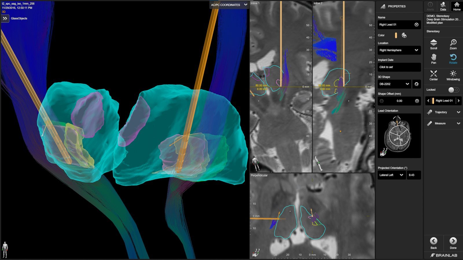
Classical targeting relies on atlas coordinates and visible landmarks on MRI. That still works for many cases, but the targets are small, deep and variable between patients. Modern workflows therefore add patient-specific information: segmentation of the nuclei that matter, outlines of structures to avoid, and white matter tracts that pass nearby.
A 2026 study in npj Parkinson's Disease from Amsterdam UMC shows how far this can go. In a prospective, non-randomized comparison, 102 patients had subthalamic nucleus targeting guided by patient-specific 7 Tesla MRI connectivity analysis, and 118 matched controls were targeted with classical anatomical landmarks. At about six months, motor improvement was 56 percent in the connectivity group and 50 percent in controls, and the share of poor responders fell from 14 percent to 4 percent. The authors are open about the limits: the design was not randomized, the control group was partly historical, the work came from a single center, and 7 Tesla imaging is not widely available.
The wider field is moving the same way. The proceedings of the 13th DBS Think Tank, published in Frontiers in Human Neuroscience in March 2026, describe tractography-guided targeting in conditions such as obsessive compulsive disorder, alongside ultrahigh-field imaging and adaptive stimulation concepts. Many of those results come from small, open-label or preliminary cohorts, and the report itself treats them that way. The direction is clear. The evidence is still maturing.
What a software planning chain contains
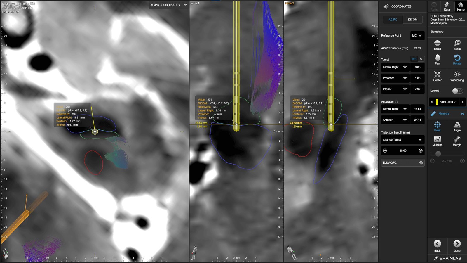
Brainlab describes its DBS solution as a chain of Elements applications. Elements Segmentation shows patient-specific anatomical structures. Elements Fibertracking creates fiber tracts from built-in or user-customizable templates. Elements Object Management segments target and avoidance structures, including those tracts. Elements Stereotaxy and Trajectory Planning Cranial support case-specific implant modeling, frame-based stereotactic localization and trajectory planning. Elements Import/Export can send the plan to third-party lead implantation systems.
The value is less in any single module than in having them in one plan. The surgeon can see the target, the tracts and the lead trajectory together, rather than moving between separate tools. Availability of individual modules differs by country, so a hospital should confirm what is cleared locally before planning around it. Our article on AI in surgical planning inside Brainlab Elements covers the automation side of the same suite.
Execution: frame, navigation or robot
There is no single right way to get the lead to the target. Frame-based stereotaxy, frameless navigation and robotic guidance are all in use, and the choice reflects the team, the volume and the hospital. A 2026 technique paper in Acta Neurochirurgica from Hospital Clinic Barcelona describes an image-based, robot-assisted workflow under general anesthesia: high-resolution MRI fused with thin-slice CT for planning, a registration accuracy check, intraoperative 3D imaging to verify the position, and the neurostimulator placed in the same session. The authors note the real constraints too: acquisition and maintenance costs, a learning curve, and the fact that the robot controls the linear trajectory but not the rotation of a directional lead, so orientation has to be checked separately.
That last point shows why imaging at the table matters. Brainlab Loop-X is a mobile intraoperative 2D and 3D imaging system that can scan a patient in a stereotactic head frame and store imaging positions for repeat control scans. It can give the team a position check while the patient is still in the room. See our overview of mobile intraoperative imaging with Loop-X, and the wider context in surgical navigation for neuro and spine.
After the lead is in: verification and programming
Directional leads add a second question after the first: not only where the lead sits, but which way its segmented contacts face. Elements Lead Localization detects the lead on postoperative CT and estimates directional lead orientation. According to Brainlab, that orientation estimate is available only for Boston Scientific directional lead models, which a hospital should keep in mind when choosing hardware.
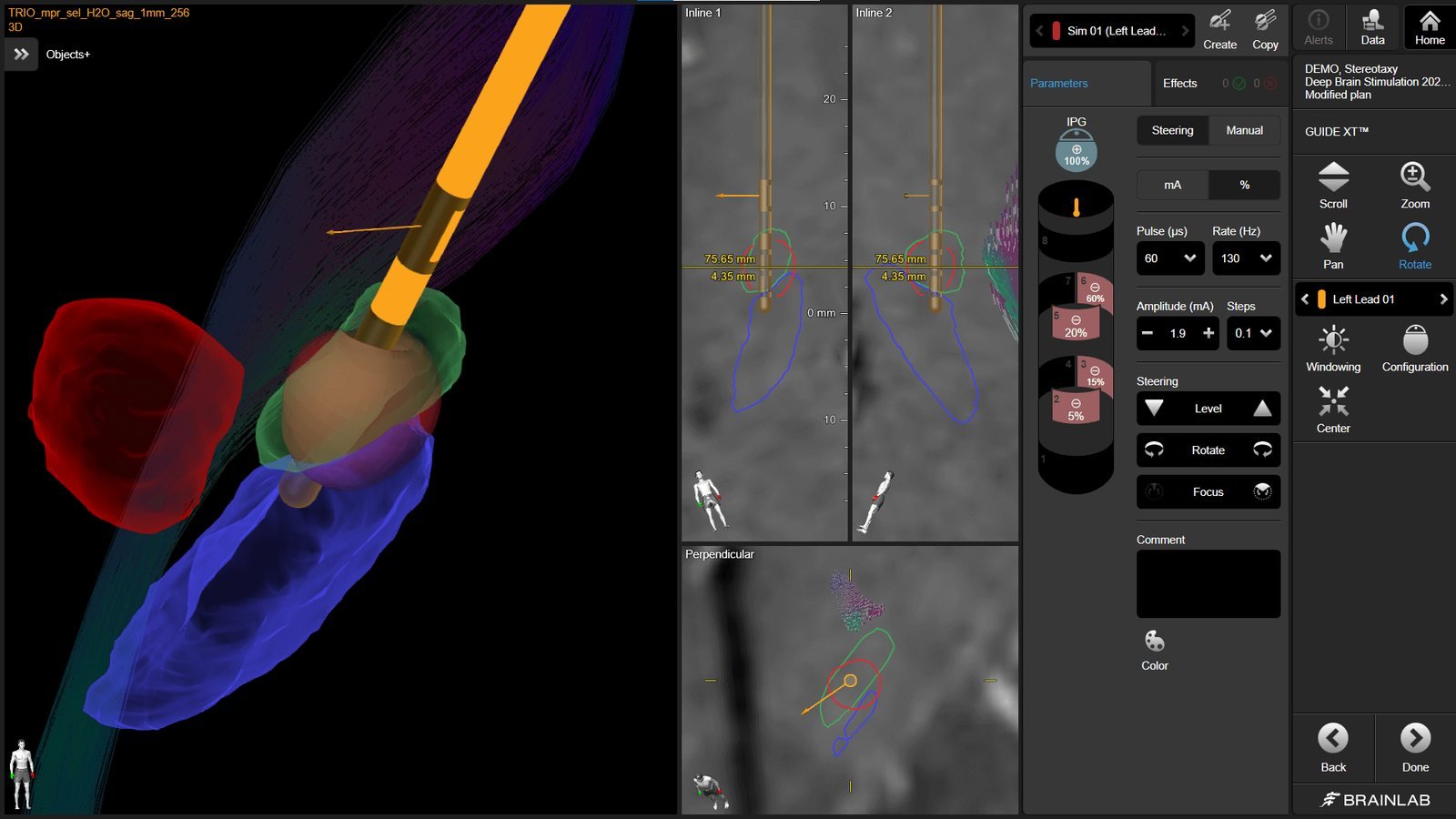
Brainlab Guide XT then shows stimulation field models in the patient anatomy, so clinicians can plan programming parameters in advance of the patient visit. Brainlab cites published work reporting reduced programming time with this approach. As with every tool of this kind, it supports the clinician's decision and does not replace it.
What a hospital needs to start or grow a DBS service
A sensible route is to build in stages. Begin with the team and the pathway: neurology, neurosurgery, neuroradiology, a neurophysiology and programming service, and clear patient selection. Add patient-specific planning so that every case is prepared consistently. Then connect execution and verification, with imaging at the table and a defined check after implantation. Each step improves the program on its own.
Brainz supports neurosurgery departments across the region with installation, training, clinical support and after-sales coordination for Brainlab platforms, and can help a team scope which pieces fit their current volume. See our neurosurgery page for the full solution area, or talk to us if you are planning a functional neurosurgery program.
Product availability, indications, regulatory status and clinical use vary by country and follow manufacturer documentation and applicable regulatory approvals. Trademarks belong to their respective owners.